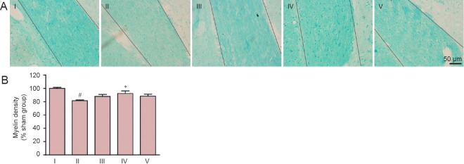
Figure 2.
Effect of carnosine on white matter damage in the optic tract in the mouse model of SIVD.
(A) Effect of carnosine on white matter damage in the optic tract was evaluated by Klüver-Barrera staining after right unilateral common carotid artery occlusion. Klüver-Barrera staining revealed white matter rarefaction 37 days after carotid occlusion. The staining intensity of the myelinated fibers was reduced in the SIVD group, and was increased by administration of carnosine (500 mg/kg). The areas between the two black lines represent the optic tract. Scale bar: 50 µm. (B) Quantitative analysis was performed using the mean optical density value (mean ± SEM). n = 8–10 mice in each group. #P < 0.05, vs. sham group; *P < 0.05, vs. SIVD group. I–V: Sham, SIVD, SIVD + carnosine 200 mg/kg, SIVD + carnosine 500 mg/kg and SIVD + carnosine 750 mg/kg groups, respectively. SIVD: Subcortical ischemic vascular dementia.

