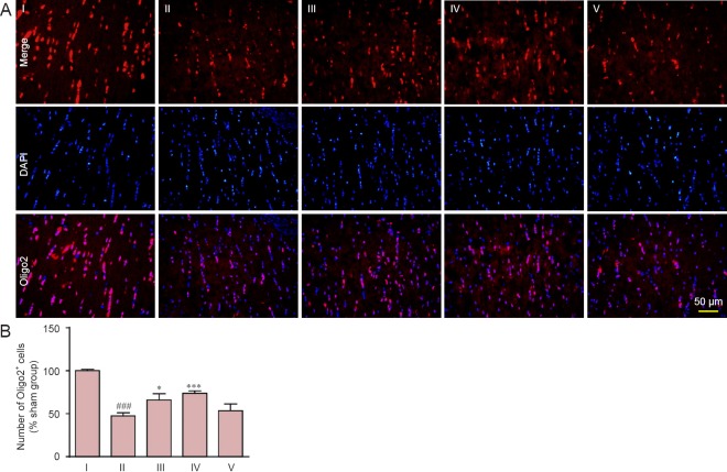
Figure 6.
Effect of carnosine on oligodendrocyte damage in the corpus callosum.
(A) The total oligodendrocyte (Oligo2+) count after carnosine treatment was calculated as the percentage of total cells (labeled by DAPI) after right unilateral common carotid artery occlusion. Fluorescent indicator is Cy3 (red). Scale bar: 50 µm. (B) Quantitative analysis of Oligo2+ cells. The numbers of oligodendrocytes (Oligo2+ cells) was decreased on day 37 after artery occlusion, and was increased by administration of carnosine (200, 500 mg/kg). Data are expressed as the mean ± SEM. n = 8–10 mice in each group. ###P < 0.001, vs. sham group; *P < 0.05, ***P < 0.001, vs. SIVD group. I–V: Sham, SIVD, SIVD + carnosine 200 mg/kg, SIVD + carnosine 500 mg/kg and SIVD + carnosine 750 mg/kg groups, respectively. SIVD: Subcortical ischemic vascular dementia; DAPI: 4′,6-diamidino-2-phenylindole.

