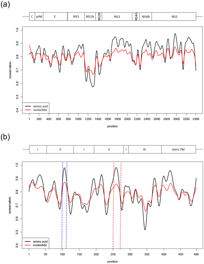
Figure 1. Genome-wide analysis of DV conserved sites.
Conservation profile of nucleotides (red line) and amino acids (black line) of 480 dengue coding sequences (CDS) (a) and their respective envelope proteins (b). The “x” axis represents the position of amino acid residues in the DV CDS (a) or Envelope protein (b) and “y” axis represents the conservation score, where 1 indicates the highest conservation. The two most highly conserved peptides in the envelope protein (b) are the fusion peptide (residues E99–112, highlighted in the blue box) and E250–270 (red box). Above “a” the schematic DV CDS. C: capsid; prM: membrane precursor; E: envelope; NS: non-structural. Above “b” the scheme of E protein domains (I, II, II), the stem segment and transmembrane anchor (TM).

