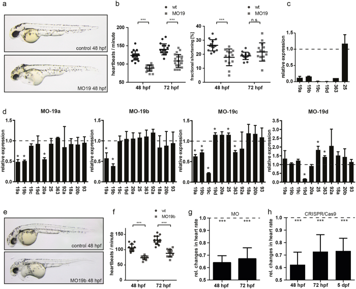
Figure 3. Loss of miR-19 leads to bradycardia.
(a) Lateral brightfield images of control-injected and MO19-injected 48 hpf zebrafish embryos. Note the cardiac edema and the blood congestion at the inflow tract developed in MO19 injected embryos. (b) Quantification of heart rate as heart beats per minute and ventricular fractional shortening as a measure for ventricular contractility in control (wt) and MO19 injected embryos. Morpholino-mediated knockdown of miR-19 in zebrafish leads to bradycardia and impaired ventricular contractility (±sd; n ≥ 14; p < 0.005). (c) qRT-PCR confirmed efficient knockdown of miR-19a-d. Notice the consistent reduction in miR-363 expression (±sd; n = 45 animals from 3 independent experiments). (d) Specific knockdown of miR-19a, miR-19c and miR-19d altered expression of neighboring miRNAs. However, knockdown of miR-19b did not interfere with expression of neighboring miRNAs (±sd; n = 45 animals from 3 independent experiments; p < 0.05). (e) 48 hpf zebrafish embryos injected with control- and MO19b-morpholino revealed that miR-19b deficiency is sufficient to mimic MO19-induced phenotype with characteristics of heart dysfunction. (f) miR-19b deficient embryos developed bradycardia with up to 36% ± 5.7% reduced heart frequency (± = sd; n ≥ 12; p < 0.005) (g,h) Relative changes in heart rate normalized to controls (dotted line) indicates that CRSIPR/Cas9 mediated knockout of miR-19b caused an identical and long-term reduction in heart rate as observed for MO19b (±sd; n = 10; p < 0.005).

