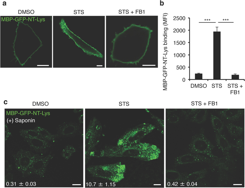
Figure 2. Staurosporine increases sphingomyelin contents in both the exofacial leaflet of the plasma membrane and intracellular compartments.
(a) Confocal images of CHO cells treated with DMSO, 50 nM STS or 50 nM STS + 15 μM fumonisin B1 (FB1) for 24 hr labelled with MBP-GFP-NT-Lysenin (Lys) protein. Bar, 10 μm. (b) Binding of MBP-GFP-NT-Lys protein to the exofacial leaflets of the plasma membrane in DMSO, STS or STS + FB1 treated CHO cells. Data are means ± SEM (n = 3). ***p < 0.001. MFI; mean fluorescence intensity. (c) Confocal images of DMSO, STS or STS + FB1 treated CHO cells stained with MBP-GFP-NT-Lys protein after membrane permeabilization by 0.05% (w/v) saponin. Bar, 10 μm. The value of fluorescence intensity of MBP-GFP-NT-Lys was shown in images. Data are means ± SEM (50 cells from three independent experiments).

