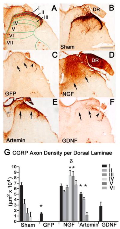
Figure 3.

Regeneration of CGRP-positive axons with neurotrophic factor expression. In normal spinal cord (A & B), CGRP+ axons are mostly located in superficial laminas I & II as indicated by the stain patterns associated with the laminae of Rexed (A). Lenti-GFP injections following L4/L5 dorsal root crush (C) did not have any effect on regeneration of CGRP+ axons past the entry zone (arrows), but showed some staining within Lissauer’s tract (arrowhead). Lentiviral expression of NGF induced robust regeneration of CGRP + axons (D), but the regeneration was ectopic and axons grew throughout most of the dorsal horn laminae (arrows). Lentiviral expression of artemin produced topographically targeted regeneration of CGRP+ fibers (E) and the axons occupied their physiological lamina (arrows). Lentiviral expression of GDNF had no effect on regeneration of CGRP+ axons (arrows) showing axons only within Lissauer’s tract (arrowhead; F). Lamina specific quantification of the area occupied by CGRP+ axons in control GFP, NGF, artemin and GDNF expressed group (G). Dorsal root injury completely abolished CGRP fibers in all laminas in the ipsilateral dorsal horn and the axon density was significantly lower in GFP lesion control compared to no lesion controls (*p<.05). Statistical analysis by a two-way ANOVA showed a significant increase in axon occupying area in lamina I and II for NGF and artemin compared to GFP controls and GDNF (*p<.05). NGF expression resulted in a significant increase in axons occupying the deeper laminae (III, IV, V and VI) when compared to the Sham or artemin groups that targeted regenerating CGRP axons to their physiological targets (lamina1&II) (δp<.05). Values represent mean±SEM, n=12 for GFP and artemin, n=9 for NGF, =7 for GDNF. Scale bar = 300 μm.
