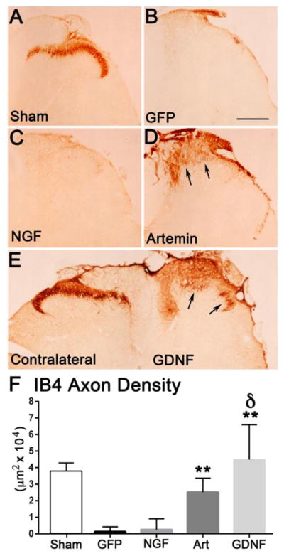
Figure 4.

Artemin and GDNF enhanced regeneration of IB4+ axons while NGF had no effect on IB4 axons. Non-injured Sham controls show a normal distribution of IB4 axons within the inner region of laminae II (A). Dorsal root crush and injections of lenti-GFP resulted in a complete abolishment of IB4+ axons and absence of regeneration (B). NGF expression in the dorsal horn did not have any effect on IB4+ axon regeneration and looked similar to GFP lesion controls (C). Artemin produced modest regeneration of IB4+ axons just past the entry zone (arrows; D). GDNF induced robust regeneration of IB4+ axons (arrows) into the appropriate laminae (right side E). Quantification of IB4+ axon occupying area within laminae I – III (F). Compared to control and NGF, artemin and GDNF produced a statistically significant regeneration of IB4+ axons (*p<.05, Tukey’s post hoc test) and the effect of GDNF was significantly higher compared to all treatment groups after dorsal root injury (δ p<.05). Scale bar = 300 μm.
