
Figure 5.
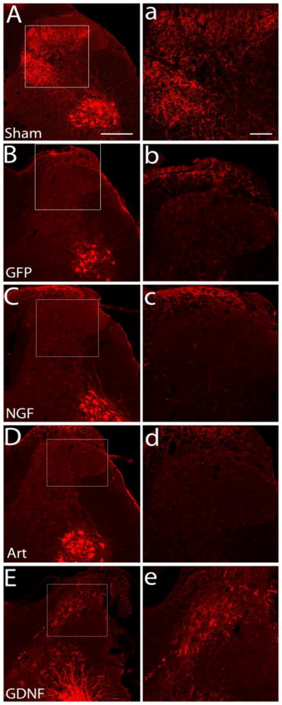
Regeneration of CTB-labeled myelinated axons after neurotrophin treatment. Sham controls show intact axons labeled by cholera toxin-B to demonstrate successful labeling and topography of the deeper myelinated afferents in a non-lesioned animal (A, a). Dorsal root crush injury completely transected all the deeper myelinated sensory afferents and there is no spontaneous regeneration of these axons in GFP control animals (B, b). Regeneration of CTB-labeled sensory afferents was absent after dorsal root injury and treatment with either NGF (C, c) or artemin (D, d). However, all animals showed very good and consistent labeling of the ventral motor neuron pools and their dendrites, indicating tracer uptake into the sciatic nerve. Many CTB-labeled sensory axons were observed to have regenerated into the spinal cord after treatment with GDNF (E, e). High magnification images (a,b,c,d, and e) are of boxed area shown in A,B,C,D, and E, respectively. n=6 for sham, GFP control and NGF, n=9 for artemin, n=7 for GDNF. Scale bar = 300μm (A,B,C,D,&E) and 100 μm (a,b,c,d, & e).