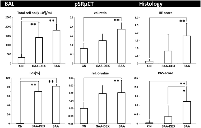
Figure 4. Correlation of XLF and UWBP to ex-vivo techniques BAL, pSRμCT, and histology for healthy mice (CN), mice with severe acute airway inflammation (SAA) and dexamethasone treated mice of the same model (SAA-DEX).
The analysis of BAL shows a significant increase in total cell number and in the relative amount of eosinophils (EOS[%]) in SAA mice when compared to CN mice, verifying the presence of asthma. SAA-DEX still reveals an increased number of eosinophils suggesting inefficient treatment. pSRμCT suggests a strongly increased soft-tissue vol.ratio within the lungs of SAA and intermediate values for SAA-DEX mice. SAA and SAA-DEX lungs show no difference in the rel. δ-value. Histology shows an increased amount of infiltrating cells in SAA (HE-score) and an increased mucus production (PAS-score), SAA-DEX mice demonstrate intermediate results, suggesting only partially successful treatment. Statistical significance difference of the results is indicated by (*p < 0.1, **p < 0.05).

