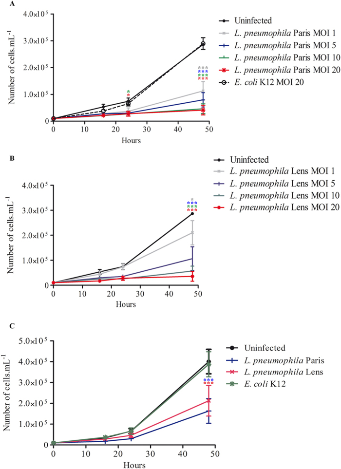
Figure 1. L. pneumophila prevents proliferation of A. castellanii.

(A) A. castellanii ATCC 30010 were infected with L. pneumophila Paris and E. coli K12 or (B) with L. pneumophila Lens at a MOI of 1, 5, 10 and 20. Infections were carried out within the PAS solution for 2 h and cells were further incubated within the PYG medium containing gentamicin. Sixteen, twenty four and forty eight hours after infection, cells were harvested for counting. Results are average of three independent experiments and errors bars represent the standard error of the mean (±SEM). (C) A. castellanii ATCC 30234 were co-cultured with L. pneumophila Paris, L. pneumophila Lens and E. coli K12 at a MOI of 20. At different time points, cells were harvested for counting. Results are average of three independents experiments and errors bars represent the standard error of the mean (±SEM). The asterisks indicate conditions that are significantly different compared to uninfected cells (*p < 0.05; ***p < 0.001).
