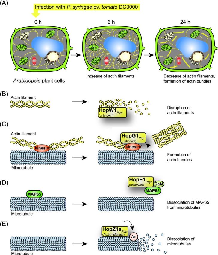
Figure 8.

Influence of type III effectors on actin filaments and microtubules. (A) Infection of Arabidopsis cells with P. syringae pv. tomato DC3000 leads to alterations in the actin cytoskeleton. The infection with wild-type or T3S mutant strains leads to an increase in actin filaments 6 hours post infection. Twenty-four hours post infection, the wild-type strain induces the formation of actin bundles and leads to a reduced number of actin filaments. Actin filaments and bundles are indicated as yellow dashes. The plant cell wall is represented in green. The following cell organelles are shown: chloroplasts (green), mitochondria (beige), vacuole (blue), nucleus (beige), ER (light brown) and Golgi apparatus (red). (B) HopW1 leads to the disruption of actin filaments. The effector protein HopW1 binds to filamentous actin and leads to the disruption of actin filaments. (C) HopG1 induces the formation of actin bundles. HopG1 binds to a mitochondrial-localized kinesin motor protein, which associates with microtubules and presumably links microtubules to actin filaments. HopG1 induces the formation of actin bundles, presumably via its interaction with kinesin. (D) HopE1 leads to the dissociation of MAP65 from microtubules. HopE1 interacts with calmodulin (CaM) and the microtubule-associated protein MAP65 and leads to the dissociation of MAP65 from microtubules. No effect of HopE1 on the microtubule network was observed. (E) HopZ1a from P. syringae dissociates microtubules. The acetyltransferase HopZ1a binds to and acetylates tubulin and disrupts microtubules.
