Figure 1.
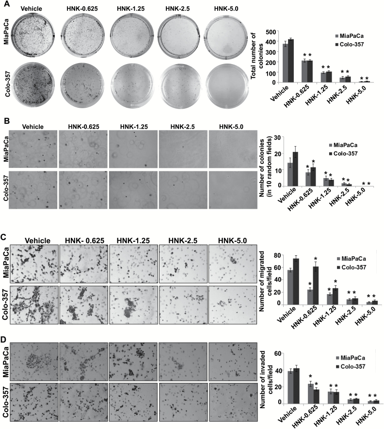
HNK inhibits growth and malignant properties of PC cells. (A) PC cells were seeded in six-well plates (1×10³ cells/well) and treated with vehicle or indicated doses of HNK. After 2 weeks of culturing, colonies were fixed, stained, photographed and quantified. (B) Equal volumes of agarose and growth medium were mixed and plated to form bottom layer in six-well plates. Cells were suspended in regular media mixed with an equal volume of agarose, and cell suspension-agar mix was seeded as top layer in each well. Cells were then incubated with vehicle or various concentrations of HNK (0.625–5.0 μM) under normal culture conditions, for 3 weeks for colony formation. Subsequently, colonies were stained with crystal violet, photographed and counted in 10 randomly selected fields. (C and D) PC cells were treated with various concentrations of HNK for 48h. Thereafter, cells were then trypsinized, counted, suspended in serum-free media and seeded equally (C) for motility [5×10⁵ (MiaPaCa) and 1×10⁶ (Colo-357) cells/well] assay on non-coated membranes and (D) for invasion on Matrigel-coated polycarbonate membrane [2.5×10⁵ (MiaPaCa) and 5×10⁵ (Colo-357) cells/well]. Media supplemented with 10% fetal bovine serum was used as chemoattractant in lower chamber. Cells were allowed to migrate/invade for 16h, and then, cells remaining in the upper portion were removed. Cells that had migrated/invaded were fixed, stained with Diff-Quick cell staining kit (Dade Behring), mounted on slides and counted in 10 random fields under microscope. Bars represent the mean ± SD (n = 3). *P < 0.05.

