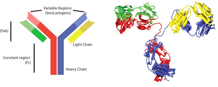
Figure 1. Antibody structure.
Antibody cartoons are often drawn in a Y-shape, as on the left. This picture represents an IgG molecule, made from two identical heavy (red and blue) and two identical light (yellow and green) chains. The top of the Y (called the Fab region) contains two variable regions that each bind the same antigen. The bottom of the Y is the constant (Fc) region, which interacts with cell receptors, complement, etc. On the right is a crystal structure of an IgG2 antibody. The heavy chains (red and yellow) and light chains (blue and green) are identical. This antibody is ∼10 nm from bottom to top. (From TimVickers. https://commons.wikimedia.org/wiki/File:Antibody_IgG2.png)

