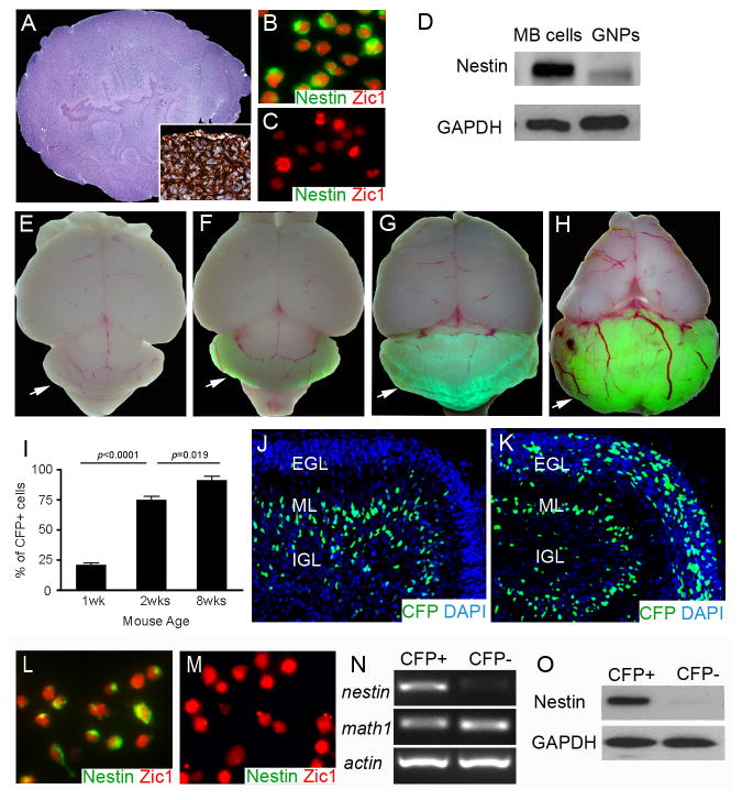
Figure 1. Cerebellar GNPs express Nestin after Ptch1 deletion.
A, Hematoxylin and eosin staining of MB from Math1-Cre/Ptch1C/C mouse at 8 weeks of age. Inset, immunostaining for Nestin in MB tissue. B–C, Immunostaining for Nestin and Zic1 on freshly isolated MB cells (B) and wildtype GNPs (C). D, Expression of Nestin and GAPDH proteins in GNPs and MB cells was examined by western blotting. e–h, Wholemount images of mouse brains from P7 Nestin-CFP mice (E), and Nestin-CFP/Math1-Cre/Ptch1C/C mice at P7 (F), P14 (G) and 8 weeks of age (H). Arrows point to mouse cerebella. I, Flow cytometry analysis of the percentage of CFP+ cells among GNPs isolated from Nestin-CFP/Math1-Cre/Ptch1C/C mice at designated stages. J–K, Immunostaining for CFP with a FITC-conjugated antibody on cerebellar sagittal sections from P7 Nestin-CFP mouse (J) and P7 Nestin-CFP/Math1-Cre/Ptch1C/C mouse (K). Cerebellar sections were counterstained with DAPI. L–M, CFP+ cells and CFP− cells were purified from cerebellar EGLs of Nestin-CFP/Math1-Cre/Ptch1C/C mice at P7 by microdissection followed by FACS. Expression of Nestin and Zic1 proteins in CFP+ cells (L) and CFP− cells (M) was examined by immunocytochemistry. N–O, mRNA expression of nestin, math1 and actin (N), and expression of Nestin and GAPDH proteins (O) in CFP+ and CFP− cells were examined by conventional RT-PCR and western blotting, respectively.

