Fig. 1.
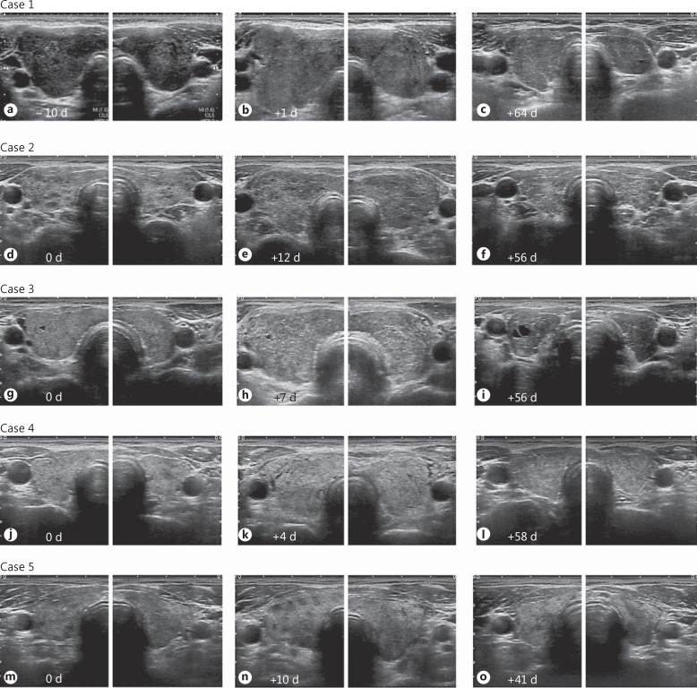
Ultrasonographic images from each case before and after 131I administration. d = Days. a-c Case 1: ultrasonographic images 10 days before (a), 1 day after (b), and 64 days after 131I administration (c). One day after neck pain developed (b), the thyroid gland was clearly enlarged and the echogenicity of the thyroid parenchyma was increased. d-f Case 2: ultrasonographic images at the same day (d), 12 days after (e), and 56 days after 131I administration (f). Three days after neck pain developed (e), a diffusely enlarged goiter with more heterogeneous internal echoes was observed. g-i Case 3: ultrasonographic images on the day (g), 7 days after (h), and 56 days after 131I administration (i). Four days after neck pain developed (h), a diffusely enlarged goiter with relatively hyperechoic internal echoes was observed. Granular hyperechoic lesions were scattered throughout the thyroid gland. j-l Case 4: ultrasonographic images on the day (j), 4 days after (k), and 58 days after 131I administration (l). One day after neck pain developed (k), a diffusely enlarged goiter was observed. m-o Case 5: ultrasonographic images at the same day (m), 10 days after (n), and 41 days after 131I administration (o). On the day the neck pain developed (n), the thyroid gland was enlarged and the echogenicity of the thyroid parenchyma was increased. The border between the thyroid gland and the surrounding tissue became blurred and irregular (cases 1-5), and the surrounding tissue was hyperechoic (cases 3-5) within several days of neck pain development (b, e, h, k, n).

