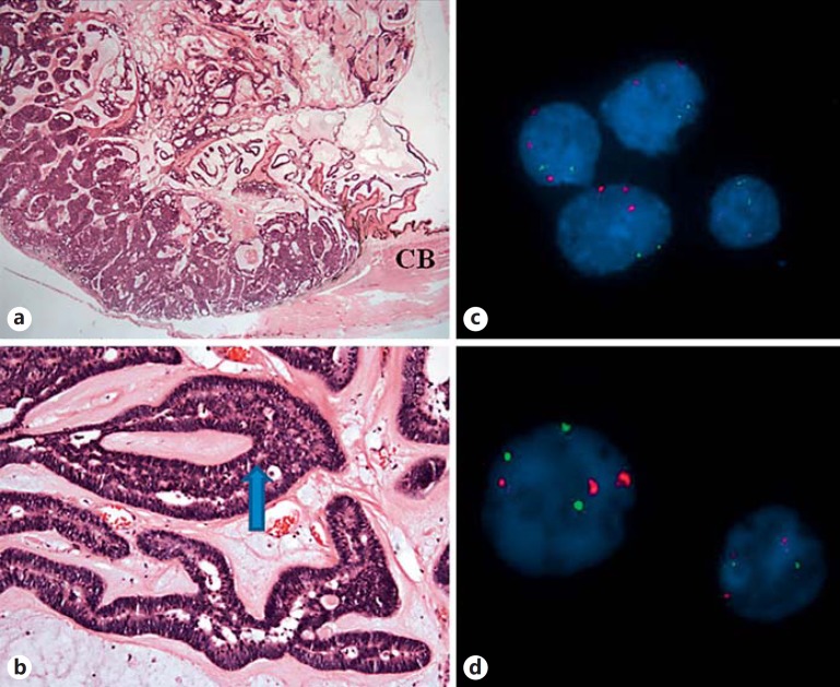
Fig. 2.
a Pale, gelatinous nodule measuring 12 × 8 mm in size. Hematoxylin and eosin. ×1. b Neuroepithelial cells forming cords, trabeculae, and rosette-like structures (arrow) reminiscent of primitive neural tube. Hematoxylin and eosin. ×10. c FISH analysis of the punch biopsy shows trisomy of chromosome 3 and the normal diploid chromosome 8. Red probe: D3Z1, centromere 3; green probe: D8Z2, centromere 8. d FISH analysis of the FNA biopsy shows trisomy of chromosome 3 and occasional trisomy of chromosome 8. Red probe: D3Z1, centromere 3; green probe: D8Z2, centromere 8. CB = Ciliary body.

