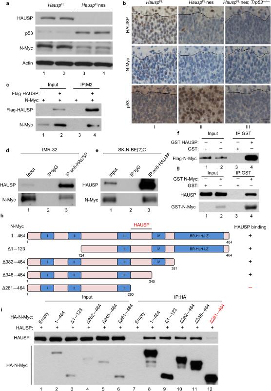
Figure 1. HAUSP affects and directly interacts with N-Myc both in vitro and in vivo.
(a) Western blot of mouse brains collected at day E13.5; n = 2. (b) Representative immunohistochemistry of HAUSP, N-Myc and p53 of E18.5 mouse cortex sections of the marginal zone to the cortical plate separated by a dotted black line; magnification 40 ×; scale bar 25 μm; n = 2 per group. (c) N-Myc expression vector cotransfected with Flag-HAUSP (lane 2) or empty vector (lane 1) in HEK293T cells. Cell lysates were incubated with Flag/M2 beads then subjected to western blot; 10% input; n = 3. (d, e) Endogenous immunoprecipitation of N-Myc with HAUSP from native IMR-32 (d) and SK-N-BE(2)C cells (e); 2% input; n = 3. (f) Direct interaction between purified Flag-N-Myc and GST-HAUSP. Purified N-Myc was incubated with GST protein (lane 1) or GST-HAUSP (lane 2) and immobilized with GST beads then subjected to western blot; 10% input; n = 2. (g) Direct interaction between purified Flag-HAUSP and GST-N-Myc as in Fig. 1f. Proteins were immobilized with GST beads then subjected to western blot; 1% input; n = 3. (h) Schematic representation of N-Myc deletion mutants used for domain mapping. (i) Indicated N-Myc expression vectors cotransfected with HAUSP in HEK293T cells. Lysates were incubated with HA beads then subjected to western blot; 10% input; n = 3.

