Figure 3.
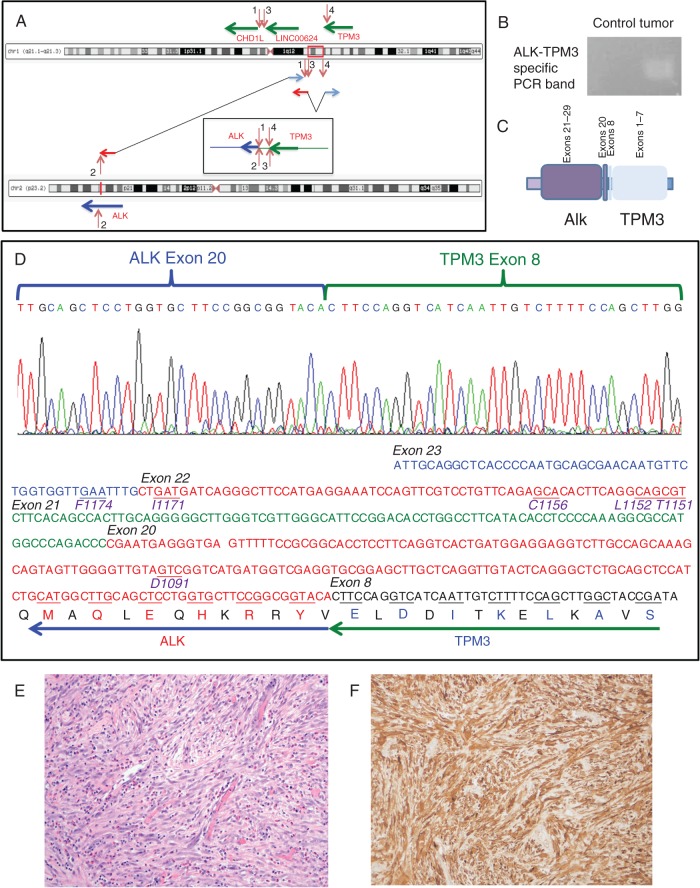
Transcriptomic and immunohistochemical analysis of TPM3–ALK rearrangement. The chromoplectic rearrangement linking a potential fusion between TPM3 and ALK is depicted (A). This rearrangement was confirmed by rtPCR in cDNA generated from tumor RNA using forward and reverse primers of exonic sequences of ALK and TPM3 (B), indicating the predicted fusion of exon 8 of TPM3 to exon 20 of ALK (C). Sanger sequencing of the PCR product confirmed this as a functional, in-frame fusion transcript. Amino acid positions of previously reported resistance mutations in the ALK kinase domain covered in the cDNA PCR amplicon sequenced are indicated in purple below the underlined codons in the DNA sequence and were demonstrated to be wild-type in this patient. (D) Histopathologic examination of the tumor revealed myofibroproliferation with increased inflammatory infiltrates including many plasma cells and eosinophils, diagnostic of inflammatory myofibroblastic tumor (hematoxylin and eosin stain, 200× original magnification) (E). Immunohistochemical staining with ALK antibody (D5F3 clone, Cell Signaling Technology) demonstrated diffuse positivity in the myofibroblastic tumor cells (200× original magnification) (F).

