
Fig. 4. 2D and 3D cell culture on architectures and within microgels with cross-scale cell patterns configured by surface properties and inner electric fields.
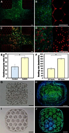
(A and B) NIH/3T3 fibroblasts, stained with Alexa Fluor 488 phalloidin (F-actin, green) and DAPI (nuclei, blue) at 48 hours in culture, growing on GelMA (8%) but not on PEGDA of the heterogeneous architecture. (C and D) Neonatal mouse cardiomyocytes patterned with GelMA/PEGDA surfaces and stained with Alexa Fluor 488 phalloidin, DAPI, and Cy3 (cTnI) at 48 hours in culture. (E) Cardiomyocyte ratio over the entire cells at 48 hours in culture on a 24-well plate as control and on GelMA of the architecture. (F) Beating rate of the cardiomyocytes at 24 and 48 hours in culture on GelMA of the architecture (movie S6). (G) GelMA (5%) microgel crosslinked with encapsulated random NIH/3T3 fibroblasts. (H) GelMA/cell microgel in (G) stained with Alexa Fluor 488 phalloidin and DAPI on day 5 in culture. (I) GelMA microgel crosslinked with encapsulated and reorganized NIH/3T3 fibroblasts (movie S7). (J) GelMA/cell microgels in (I) stained on day 3 in culture. *P < 0.01. Scale bars, 200 μm.