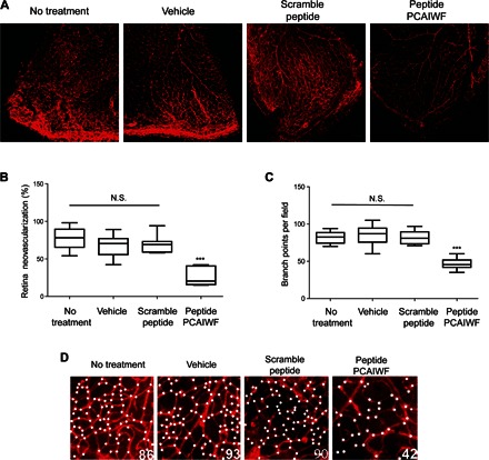
Fig. 7. PCAIWF inhibits neovascularization in vivo.

Neonatal C57BL/6 mice with OIR at P15 were treated or not treated (n = 9 retinas) with a single intravitreal injection (1 μl) of vehicle only [dimethyl sulfoxide (DMSO)] (n = 11 retinas), peptide PCAIWF (30 μg) (n = 6 retinas), or scramble peptide IFCAPW (30 μg) (n = 7 retinas). Whole-mount retinas were stained with isolectin-B4 conjugated to Alexa Fluor 594 red-fluorescent dye. (A) Representative confocal microscopy images of retinas of OIR neonatal C57Bl/6 mice at P17. (B) Quantification of neovascularization in the retinas of OIR neonatal C57BL/6 mice treated or not treated with peptides. (C) High-magnification images (×200) of the retinas at P17 of OIR neonatal C57BL/6 mice treated or not treated with peptides. White dots indicate vessel sprouts or bifurcations. The numbers of sprouts/bifurcations determined for each image are indicated at the bottom right corner. (D) Quantification of vessel sprout or bifurcations. Statistics, ANOVA (Tukey’s multiple comparison test) (N.S., P > 0.05; ***P < 0.005). Box plots in which the boxes define the 25th and 75th percentiles, with a line at a median and error bars defining the 10th and 90th percentiles.
