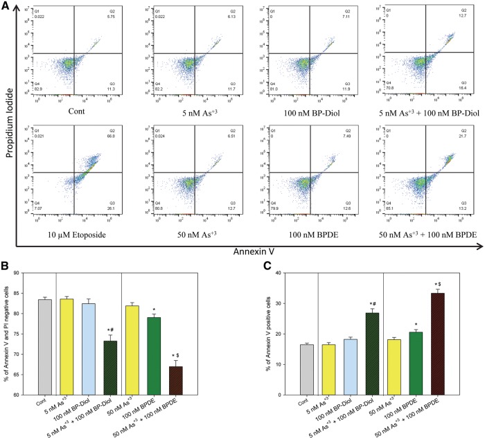
FIG. 3.
Annexin V and Propidium Iodide staining in primary thymus cells treated with As+3, BP-diol/BPDE and the combinations in vitro. Primary thymus cells isolated from C57BL/6J male mice were exposed to 5 or 50 nM As+3, 100 nM BP-diol or BPDE and the combinations of As+3 and BP-diol/BPDE for 18 h in vitro. A. flow cytometry results showing cells which are Annexin V-PI- (LL), Annexin V+PI- (LR), Annexin V-PI+ (UL), or Annexin V+PI+ (UR). B, viability (% of Annexin V-PI- cells). C. % of early and late apoptotic cells (% of Annexin V+ cells). *Significantly different compared to control (p < 0.05). # Synergistic effect compared to 5 nM As+3 and 100 nM BP-diol (CDI > 1). $ Synergistic effect compared to 50 nM As+3 and 100 nM BPDE (CDI > 1). Results are Means ± SD.

