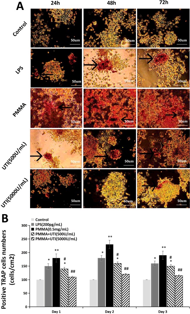
Figure 2. UTI suppressed the expression of TRAP in LPS/PMMA-induced Raw264.7 cells in a dose-dependent manner.
The cells were stimulated with alone LPS (200 pg/ml) or treated with or without various concentration (500 and 5000 units/ml) of UTI for 3 h prior to being treated with 0.5 mg/ml PMMA with adherent LPS for 24, 48 and 72 h. (A) Representative TRAP staining images of Raw264.7 cells from each group. The presence of dark purple staining granules in the cytoplasm was determined as the specific criterion for TRAP-positive cells (the black arrow points). (B) TRAP-positive cells number (cells/cm2) shown in (A) was quantified by pixel area count. Data are presented as mean±S.E.M. from three independent experiments. *, P<0.05 and **, P<0.01 compared with control; #, P<0.05 and ##, P<0.01 compared with PMMA group.

