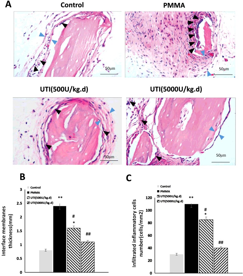
Figure 6. Total thickness of interfacial membrane (mm) and infiltrated inflammatory cells (cells/mm2) both were decreased by UTI in a dose-dependent manner in PMMA-induced murine osteolysis model.
The animal models were treated with or without various concentration of UTI (500 or 5000 units/kg i.p.) for 3 h prior to PMMA with adherent LPS implantation, and then UTI (500 or 5000 units/kg per day) was continuously injected (i.p.) once per day for 21 days. (A) Representative HE staining images of distal femur from each group. Total thickness of interfacial membrane (mm) and infiltrated inflammatory cells are indicated by blue and black arrowheads respectively. (B) Total thickness (mm) and (C) infiltrated inflammatory cells (cells/mm2) were quantified using automated image analyser. All values are expressed as mean ± S.E.M. n=10 mice per group. *, P<0.05 and **, P< 0.01 compared with control; #, P<0.05 and ##, P<0.01 compared with PMMA group.

