Figure 3.

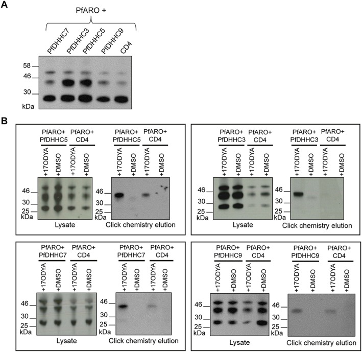
Palmitoyl‐transferase activity assay demonstrating the palmitoyl‐transferase activity of the PfDHHC proteins on PfARO. A. Immunoprecipitation of PfARO co‐expressed with each PfDHHC protein. Human embryonic kidney 293E cells were co‐transfected with plasmids coding for the expression of c‐Myc‐tagged PfARO, along with the indicated FLAG‐tagged PfDHHC proteins (PfDHHC3, 5, 7 and 9) or the control vector (CD4). PfARO was immunoprecipitated from cell lysates using α‐c‐Myc antibody. The proteins were separated by SDS‐PAGE and visualized by immunoblot, using α‐c‐Myc antibody from a different species. B. PAT activity assay. Human embryonic kidney 293E cells were co‐transfected with plasmids expressing c‐Myc‐tagged PfARO, along with the indicated FLAG‐tagged PfDHHC protein or the control vector, CD4. Cells were either treated with the metabolic label, 17‐octadecynoic acid or mock‐treated with DMSO. Proteins were extracted, and an aliquot of each lysate kept aside to confirm protein expression. The remaining lysates were put through click chemistry reactions to biotin‐azide, and 17‐octadecynoic acid‐labelled proteins were streptavidin affinity purified and eluted by boiling in SDS. Samples from the initial lysates and the click chemistry elutions were separated by SDS‐PAGE, and the presence of c‐Myc‐tagged PfARO in each of the samples was observed by immunoblot using antibodies against the c‐Myc tag.
