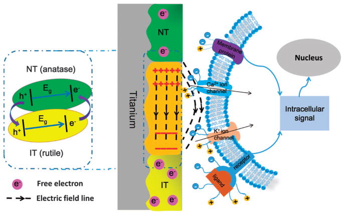
Figure 1.
Illustration of the mechanism used to generate the microscale electrostatic field (MEF) and the interaction between the MEF and the stem cells. (Left) The MEF on the titanium surface. NT refers to the non-irradiated n-type semiconducting anatase TiO2 zone prepared by hydrothermal synthesis, and IT refers to the n-type semiconducting rutile TiO2 zone prepared by laser irradiation of the anatase TiO2 zone. The MEF was generated by the surface potential difference between the NT and IT zones. The diagram in the dashed line box illustrates the TiO2 phase junction of the NT and IT, as well as the electron transfer from the NT (rutile) to the IT (anatase) zone.21,42 (Right) Stem cell membrane with charged protein affected by MEF. As ion channels, membrane proteins, ligands and receptors are all charged with different surface potentials, their surface charges would be polarized under the guidance of MEF. The sustained built-in MEF enables the polarized stem cell surface species to transduce signals to the nucleus to activate osteogenic genes that results in enhanced osteogenesis.

