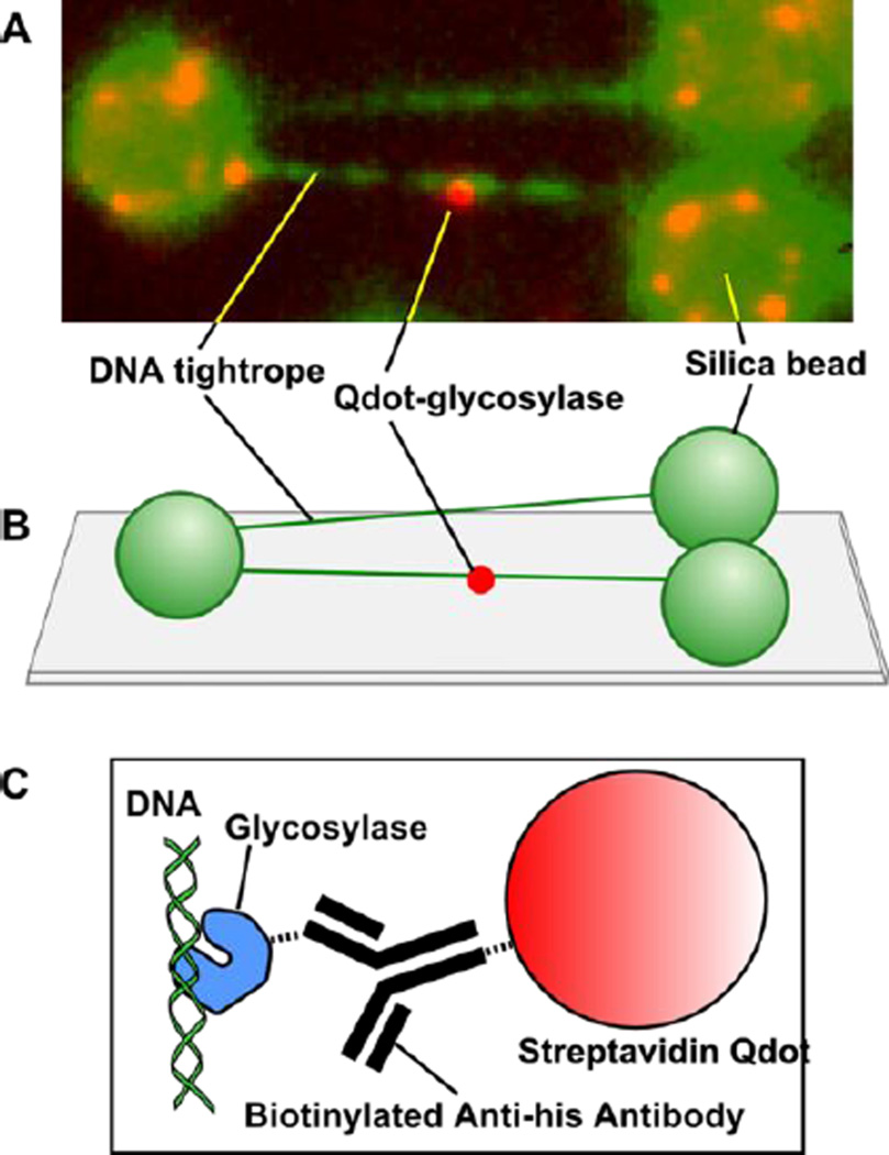
Figure 2. Single molecule sample configuration.
(A) Image of a single Qdot-labeled glycosylase scanning along elongated λ-DNA. (B) λ-DNA tightropes are elongated between polylysine-coated silica spheres on a PEG-coated microscope coverslip. (C) The 6-his tagged glycosylase is conjugated to a streptavidin Qdot through a biotinylated anti-his antibody.

