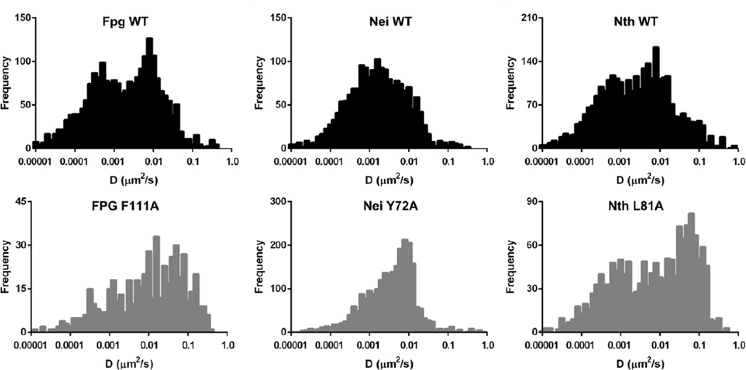
Figure 7. Time-weighted diffusive behavior of wild-type and wedge variant glycosylases scanning along damaged λ-DNA.
Fpg WT and F111A and Nth WT and L81A data are shown at low doses of damage (approximately 100 damages per λ molecule). Nei WT and Y72A are shown at a high dose of damage (approximately 280 lesions per λ). See (Nelson et al., 2014) for further details of experimental conditions.

