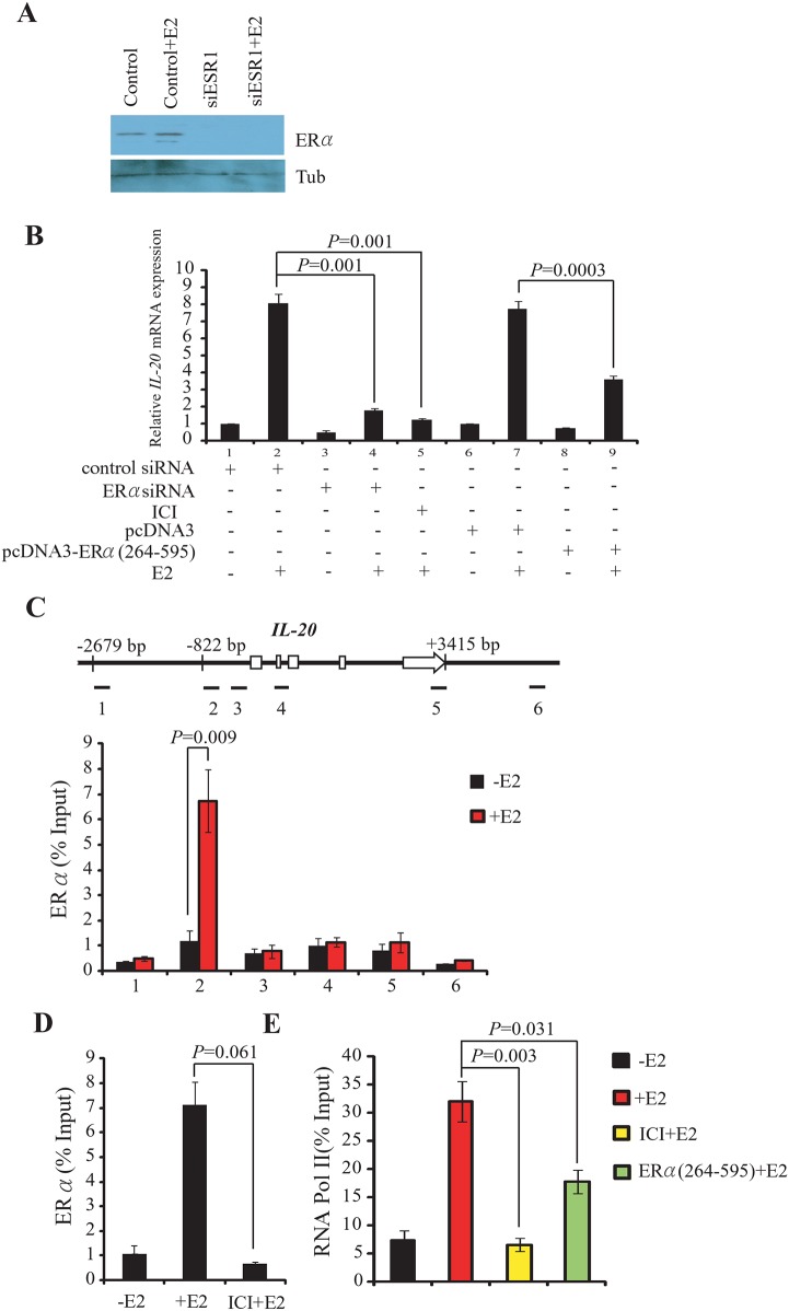
Fig 2. ERα is required for the E2-mediated induction of IL-20.
(A) Western blot shows the protein level of ERα in cells transfected with the siESR1 following E2-stimulation for 30 min. (B) Expression of IL-20 in MCF-7 cells is dependent on the presence and activity of ERα and E2, and normalized against 18s rRNA. (C) Binding of ERα to the IL-20 promoter region determined by ChIP assays. Upper panel: Schematic of the IL-20 locus (exons as open boxes) and the six amplicons (black segments) used in ChIP assays. The specific anti-ERα antibody, HC-20X, was used in the ChIP experiments. Lower panel: Bar chart of the relative levels of ERα at each of the six regions. The mean and SD were calculated from at least two independent experiments. (D) Suppressed ERα binding to the IL-20 promoter (segment 2) by ICI treatment. The ChIP experiment was carried out in the absence or presence of E2 for 30 min. (E) Inhibition of RNA Pol II binding to the IL-20 promoter (segment 2) by ICI treatment or over-expression of truncated ERα (ERα264–595) determined by ChIP. The experiment was carried out in the presence or absence of E2 for 30 min.

