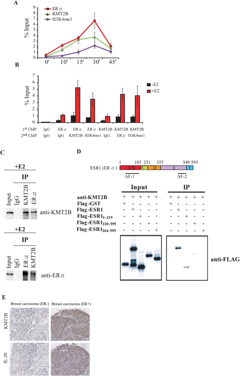
Fig 5. KMT2B and ERα form a complex at the promoter of the IL-20 gene.
(A) Kinetic ChIP experiments were performed using KMT2B, H3K4me1, and ERα specific antibodies. A single chromatin was prepared for ChIP assay at each time point. (B) ChIP-ReChIP to determine the KMT2B and ERα co-occupancy at the IL-20 promoter. Chromatin was prepared from MCF-7 cells treated with E2 for 30 minutes and then subjected to the ChIP procedure using the antibodies labeled as "1st ChIP.” The second immunoprecipitation was carried out using the antibodies labeled as "2nd ChIP.” (C) Co-immunoprecipitation of endogenous KMT2B and ERα. MCF-7 cells were treated with E2 for 24 h, and whole-cell lysates were immunoprecipitated using KMT2B or ERα antibodies. Western blotting was performed on the immunoprecipitated proteins using anti-KMT2B or anti-ERα. (D) Upper panel: Schematic of the ERα functional domains. Lower panel: Immunoprecipitation analysis of the ERα functional domains that interact with KMT2B. Interactions between the endogenous KMT2B and the in vivo transcribed/translated Flag-tagged ERα fragment were confirmed by an immunoprecipitation assay using anti-KMT2B antibody followed by western blotting with anti-FLAG antibody. (E) KMT2B and IL-20 protein expression in ER-positive and ER-negative breast cancer assayed by IHC.

