
Figure 3.
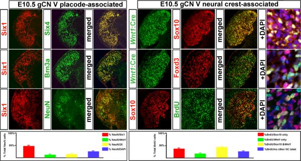
Placode versus neural crest signatures of distinct cranial sensory cell classes. The data presented here focuses on the g CN V; however, similar observations were made in the additional cranial ganglia. Left: The identity and early neuronal differentiation of cells expressing placode-associated markers. At left, cells immunolabeled with Six1 antisera (left panel, left column) to identify placode-associated cells were double-labeled with Six4 (middle column, top) or Brn3a (middle column, center), or the neuronal marker NeuN (middle column, bottom). Both Six4 and Brn3a show nearly complete identity with the Six1-lableled cells (right column, merged). Many of the NeuN labeled cells are co-labeled with Six1 (right column, bottom). At bottom left, a quantitative summary of placode-associated versus neural crest-associated labeling of g CN V cells. Right: distinct, neural crest-associated molecular signatures of Wnt1:Cre recombined and “DAPI-only” cells in g CN V. There is some overlap in the Wnt1:Cre recombined and Sox10-expressing cells (top row); however it is far from complete. In addition to the Wnt1:Cre/Sox10 labeled cells (yellow) there are several Sox10/DAPI-only cells (violet) as well as some cells that are labeled for neither neural crest-associated marker (far right, top). There is even less overlap between the Wnt1:Cre population and the Foxd3 labeled cells (middle row). There are relatively few Wnt1:Cre recombined cells that are labeled by Foxd3 (yellow; far right); however, the DAPI-only cells labeled by Foxd3 (are fairly frequent (violet; far right). BrdU is incorporated in Sox10-expressing cells (bottom panels). The overlap is extensive (yellow cells at far right); however, there also are non-labeled cells that are labeled by BrdU. At bottom, a quantitative summary of BrdU labeling seen in cells that express neural crest versus non-neural crest markers.