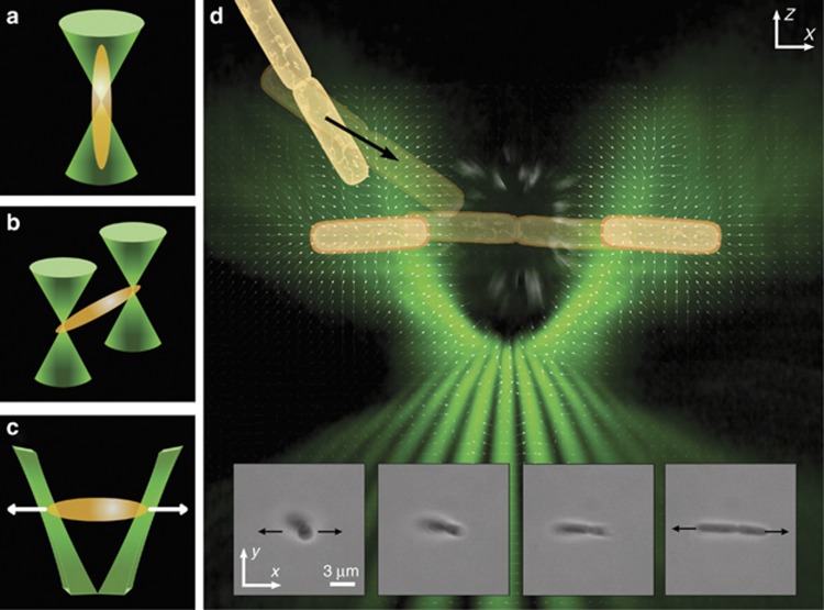
Figure 1.
Different designs of optical tweezers (left panels). (a) Single-beam optical tweezers tend to align a rod-shaped bacterium along the beam axis. (b) Dual-beam optical tweezers trap a bacterium at each end to orient the cell into the observing plane. (c) TOW optical tweezers trap a bacterium at each end but also exert lateral forces in opposite directions. Composite image illustrating the process of how a bacterial cluster is trapped, stretched and separated by the TOW tweezers (right panel). The diagram in (d) comprises the vector field of the intensity gradient of the trapping beam (white arrows), a volumetric rendering of the beam from experimental data near the focus of an objective lens (green shading), and a schematic representation of a pair of attached bacterial cells being trapped and pulled apart. The inserts in d show snapshots of a dividing B. thuringiensis cell that was aligned gradually onto the observing plane and stretched by the TOW tweezers (Supplementary Movie 1).

