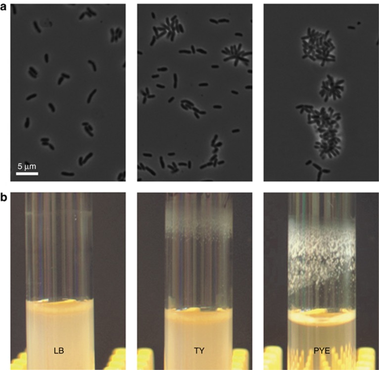
Figure 2.
S. meliloti podJ+ cells display distinct assemblages, as well as different levels of flocculation and biofilm formation, in three growth media (LB, TY and PYE). Top panels (a) show phase contrast images of the S. meliloti cells and aggregates, while bottom panels (b) show photographs of corresponding bacterial biofilms formed in 16-mm-diameter glass tubes.

