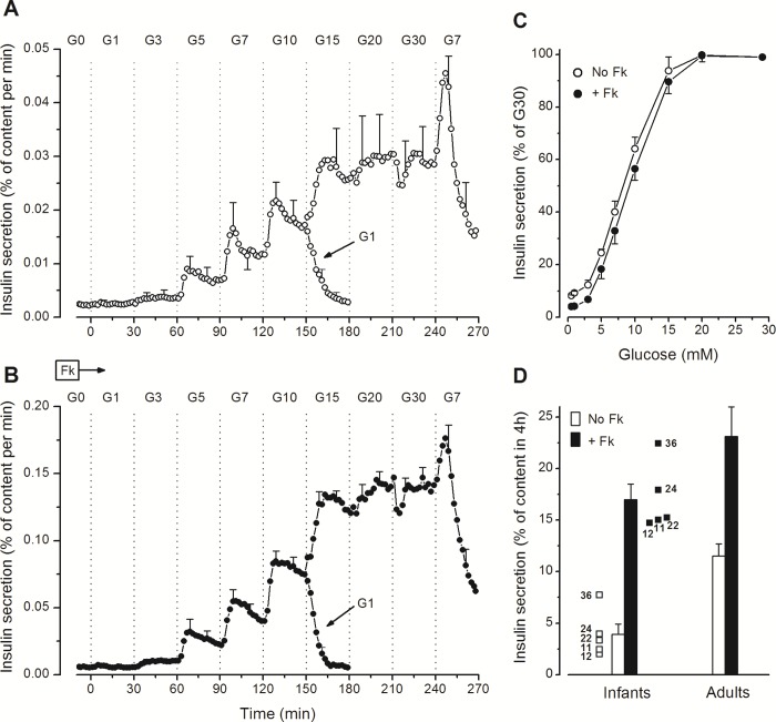
Fig 1. Concentration-dependency of glucose-induced insulin secretion in perifused islets from human infants.
(A and B) The concentration of glucose (G in mmol/l) was increased and decreased as indicated, but the islets were not exposed to the whole range of concentrations. One group of islets was perifused in G0 for 60 min before the glucose concentration was increased stepwise to G10 and eventually decreased to G1 at 150 min. Another group from the same preparation was perifused in G7 for 60 min before the glucose concentration was increased stepwise to G30 and eventually decreased to G7. Insulin secretion rates in G7 and G10 from the two series, run in parallel, were similar and therefore averaged to obtain the full dose curve for each of the five islet preparations. Parallel experiments were done in the absence (A) or presence (B) of 1 μmol/l forskolin (Fk) in islets from the five infants. (C) Concentration-dependency curves expressed as percentages of insulin secretion rates in G30. Values are means ± SE for the five infant cases. (D) Total insulin secretion (without and with forskolin) was calculated between 0 and 240 min and is shown for each of the five infant cases identified by their age in months. Columns show means ± SE for the five infant cases and for previously reported results with 8 preparations of adult islets [33].

