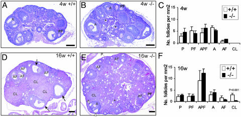
Fig. 2.
Morphological comparison of ovaries from AR+/+ and AR–/– females. (A and B) Ovaries from 4-week-old AR+/+ and AR–/– females were histologically similar. (C) Statistical analysis of the number of the follicular compartments in AR+/+ and AR–/– ovaries (n = 2). (D and E) The ovaries from sexually mature, 16-week-old AR+/+ females, compared with their AR–/– counterparts. The granulosa layers in the antral follicles were locally thin (open arrowheads) in the AR–/– ovaries, whereas they were even in thickness in the AR+/+ ovaries (arrows). An asterisk marks the zona pellucida remnants. (F) Statistical analysis of the number of the follicular compartments in AR+/+ and AR–/– ovaries (n = 3 for each genotype). Statistical significance determined by using Student's unpaired and two-tailed t test is indicated. Representative sections are shown. P, primordial and primary follicle; PF, preantral follicle; APF, atretic primordial, primary, and preantral follicle; A, antral follicle; AF, atretic antral follicle; CL, corpus luteum. (Bar: 200 μm.)

