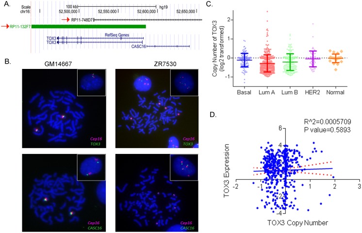
Fig 7. Copy number of TOX3 in breast tumors.
(A) Genomic positions of BAC RP11-132F7 and RP11-748D7 at chromosome 16q12. These clones were selected for homebrewed TOX3 and CASC16 FISH probes, respectively. (B) Representative FISH photomicrographs of TOX3:CEP16 and CASC16:CEP16 in GM14667 (control normal lymphocytes) and ZR-75-30 (luminal) cell lines. Cells were counterstained with DAPI (blue), while TOX3 or CASC16 is localized by green fluorescent signal, and CEP16 is localized by a red fluorescent signal. Metaphase and interphase (insert) cells are shown. These results are summarized in S2 Table. (C) TCGA copy number data analysis shows copy number of TOX3 in breast cancer subtypes. (D) Scatter plot shows no correlation between copy number and expression of TOX3 in the breast tumors.

