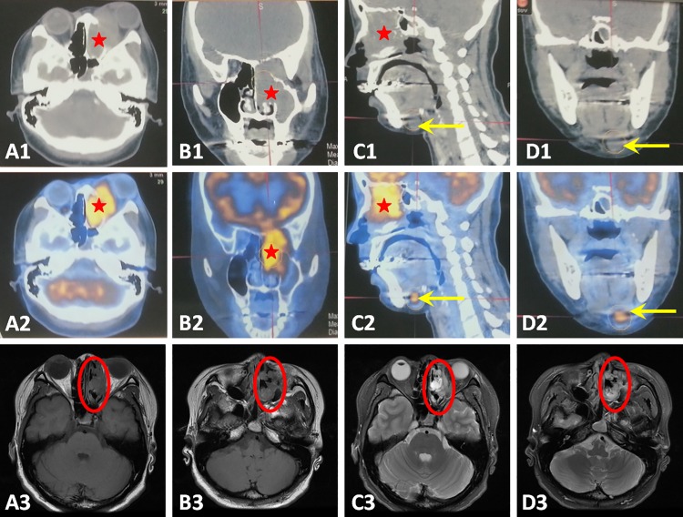
Fig 1. CT scans, PET scans and MRI findings of the tumor and metastatic cervical lymph nodes.
The tumor was seen in the left nasal cavity, maxillary and ethmoidal sinus on CT scans (A1, B1and C1, red stars) and PET scans (A2, B2 and C2, red stars). MRI T1-weighted image showed a low-intense mass (A3 and B3, red circles) while T2-weighted image showed a relatively high-intense mass (C3 and D3, red circles). Metastatic cervical lymph nodes can been seen on CT scans (C1 and D1, yellow arrows) but more prominent on the PET scans (C2 and D2, yellow arrows).

