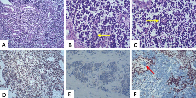
Fig 3. Histopathology characteristics of ONB.
H-E staining revealed a nest-like or cord-like tumor mass, showing uniform small cells with prominent round nuclei and eosinophilic fibrillary background (A, ×100). Pseudorosette formation (Homer-Wright rosettes) consisted of a ring of columnar cells and the presence of fibrillary material within the central space (B and C, yellow arrow, ×400). IHC staining for Ki-67 was 60% positive in all neoplastic cells (D, ×100) while staining for NSE was mild positive (E, ×400). LCA (CD45) was negative in ONB tissue but positive in lymphoid tissue (F, ×100). Tumor cells were metastatic into the lymphatic vessel (F, red arrow, ×100).

