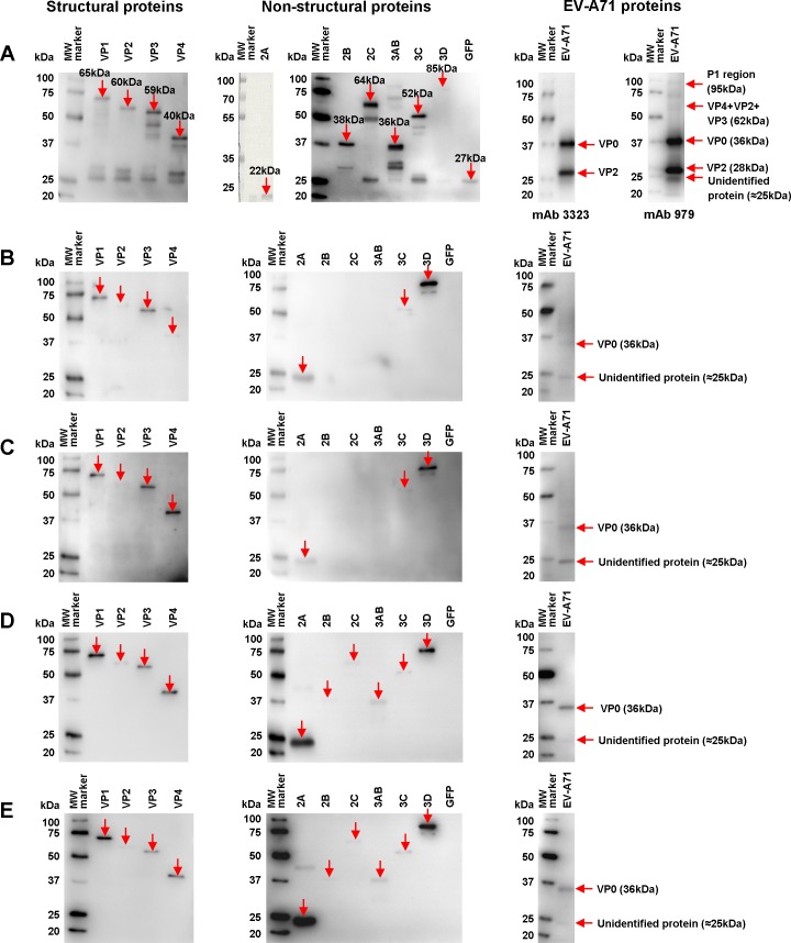
Fig 1. Antigenic profiles of the human anti-EV-A71 antibodies.
(A) Control cell lysates were loaded into SDS-PAGE gel electrophoresis. Recombinant EV-A71-EGFP cell lysates (structural and non-structural proteins) were probed with anti-GFP-HRP, while recombinant EV-A71 2A cell lysates were stained with Coomassie brilliant blue R-250. EV-A71 virion proteins were immunodetected with EV-A71-specific mAb 3323 (Millipore, USA) and mAb 979 (Millipore, USA), followed by secondary anti-mouse IgG-HRP. The expected band for each individual recombinant protein is indicated by red solid arrows and the protein sizes are shown. (B) Acute infection with no neutralization sera (n = 2) and (C) acute infection with high neutralization sera (n = 12) were used for EV-A71-specific IgM antibody detection. (D) Acute infection with high neutralization sera (n = 12) and (E) convalescent sera (n = 5) were used for EV-A71-specific IgG antibody detection. An estimated 20 μg of proteins was loaded for SDS-PAGE gel electrophoresis. The amount of EV-A71 structural and non-structural protein cell lysates was normalized with anti-GFP-HRP since the presence of inhibitory factors affected accurate quantitation of total proteins. The EV-A71 protein cell lysates and EV-A71 proteins were subjected to SDS-PAGE gel electrophoresis and probed with pooled human sera at a dilution of 1:300. The immunoblot was developed with Clarity Western ECL substrate and detected by chemiluminescence. Protein bands were determined using the Precision Plus Protein WesternC Standard (Bio-Rad, USA). The antigens recognized by EV-A71-infected patient sera are indicated by red solid arrows.

