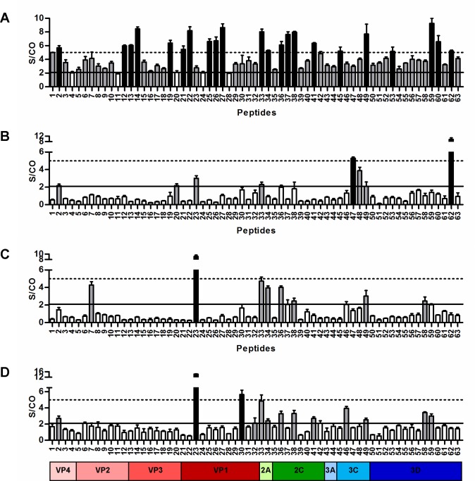
Fig 2. Mapping of EV-A71 B-cell epitopes.
Pooled human sera, at optimized dilutions of 1:2000 (IgM) and 1:500 (IgG), were subjected to peptide-based ELISA. (A) Acute infection with high neutralization sera (n = 5) were used for EV-A71-reactive IgM antibody detection. (B) Acute infection with high neutralization sera (n = 5), (C) convalescent sera (n = 3), and (D) adult sera (n = 5) were used for EV-A71-reactive IgG antibody detection. Non-HFMD children sera (n = 4) were used as negative controls. Data are presented as mean ± SD of 3 replicates. Values above the solid black line (S/CO≥2.1) were scored as weakly positive and values above the dotted line (S/CO≥5) were scored as strongly positive reactions. Grey bars represent weakly positive human anti-EV-A71 epitopes and black bars represent strongly positive human anti-EV-A71 epitopes.

