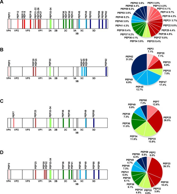
Fig 3. Analysis of anti-EV-A71 antibodies recognizing linear B-cell epitopes.
(A) IgM antibody determinants identified from acute infection with high neutralization sera. IgG antibody determinants identified from (B) acute infection with high neutralization sera, (C) convalescent sera, and (D) adult sera. Regions of amino acid sequences corresponding to the identified B-cell epitopes are indicated in the schematic diagrams of the EV-A71 genome. The percentage of antibody recognition contributed by each individual EV-A71 epitope is indicated in the pie charts, and was calculated according to the following equation: % antibody recognition = 100 x (OD values from individual peptide group/sum of the OD values from all peptide groups). In this calculation, the avidity and affinity of the peptides to the sera were assumed to be similar. Peptides are colour-coded according to the respective viral proteins.

