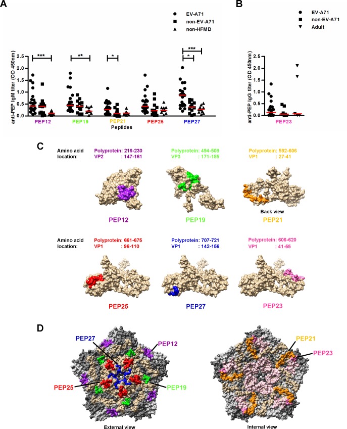
Fig 5. EV-A71-specific IgM and IgG antibody determinants.
(A) EV-A71-specific IgM antibody detection in sera (n = 44) at a dilution of 1:2000 was determined by peptide-based ELISA. Sera were categorized into EV-A71-infected patients (n = 22), non-EV-A71 enterovirus-infected patients (n = 12) and non-HFMD patients (n = 10). Red solid lines represent medians. (B) EV-A71-specific IgG antibody detection in sera (n = 38) at a dilution of 1:500 was determined by peptide-based ELISA. Sera were categorized into EV-A71-infected patients (n = 25), non-EV-A71 enterovirus-infected patients (n = 8) and healthy adults (n = 5). Red solid lines represent medians. One-way ANOVA with Kruskal-Wallis test was used for statistical analysis (*P<0.05, **P<0.01, ***P<0.001). (C) Schematic representation of locations of IgM and IgG antibody determinants in VP1, VP2 and VP3 proteins, based on structural data retrieved from PDB records (identifier 3VBS). (D) The EV-A71 pentamer structure (identifier 3VBS) was generated using UCSF Chimera software. The capsid proteins of EV-A71 are shown in brown (VP1), light grey (VP2), dark grey (VP3) and light pink (VP4). The IgM and IgG antibody determinants are indicated in purple (PEP12), green (PEP19), orange (PEP21), red (PEP25), blue (PEP27) and pink (PEP23).

