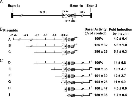
Fig. 1.
Activation of mouse SREBP-1c promoter in rat primary hepatocytes. (A) Schematic of partial mouse Srebp-1c gene, illustrating the upstream regulatory elements and the intron between exons 1c and 2. The proximal promoter regulatory region is shown with its cis-acting elements: two LXREs (open boxes) and one NF-Y and one SRE (hatched boxes). (B and C) Deletion analysis of SREBP-1c promoter region and its effects on basal and insulin-induced luciferase activities. The indicated plasmids were transfected into primary hepatocytes as described in Methods. Six hours after transfection, the medium was switched to medium B with or without 100 nM insulin and incubated for 21 h, after which the cells were harvested and assayed for dual luciferase activities as described in Methods. The 100% values for basal activity correspond to the normalized luciferase activity obtained with plasmid A (B) or plasmid D (C) in the absence of insulin. The fold induction in B and C was calculated as the ratio of normalized luciferase activity in the presence of insulin to that in the absence of insulin. Each value represents the mean ± SEM of three independent transfection experiments (each assayed in duplicate).

