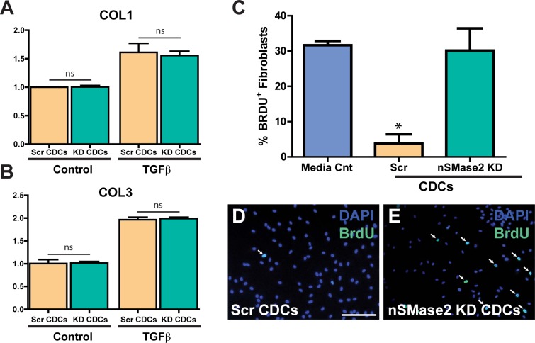
Fig 8. hCDC exosomes reduce proliferation of cardiac fibroblasts without affecting collagen gene expression.
(A, B) Human quiesced cardiac fibroblasts cocultured with either Scr CDCs or KD CDCs showed no statistically significant difference in COL1 or COL3 gene expression by qRT-PCR following TGFβ stimulation (n = 3). Data normalized to unstimulated Scr CDCs. (C, D) Cardiac fibroblasts cocultured with Scr CDCs for 24 hours showed a significant reduction in cellular proliferation as assessed by BrdU staining (5 hour pulse) when compared with (C, E) cardiac fibroblasts cocultured with nSMase2 KD CDCs and media control. n = 3. Scale bar 200μm. *p<0.05 using one-way AVOVA (Tukey’s post hoc test). Data are presented as mean ± SEM. White arrows highlight cells that co-stain for DAPI and BrdU.

