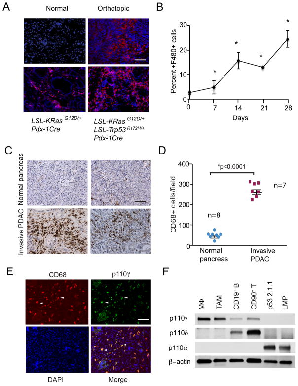
Figure 1. PI3Kγ is a marker of pancreatic ductal adenocarcinoma associated macrophages.
A. Immunofluorescent staining of F4/80+ macrophages (red) counterstained with DAPI (blue) in pancreata from normal mice, LSL-KRasG12D; Pdx-1Cre (KC) mice, LSL-KRasG12D/+; LSL-Trp53R172H/+; Pdx-1Cre (KPC) mice and mice that were implanted orthotopically with LSL-KRasG12D/+; LSL-Trp53R172H/+Pdx-1Cre (Orthotopic) tumors. Bar indicates 50μm. B. Quantification of the increase in F4/80+ macrophages over time in pancreata that were orthotopically implanted with LMP tumor cells (n=10), *p<0.01. C. Representative histopathologic characterization of human invasive pancreatic ductal adenocarcinoma tissues (n=7) and normal pancreas (n=8) showing immune reactivity for the macrophage marker CD68, with hematoxylin counterstaining. Scale bar indicates 100μm. D. Quantification of CD68+ macrophages/100X microscopic field in tissue sections from normal human pancreata (n=7) and from invasive pancreatic ductal adenocarcinomas (n=8). Statistical significance was determined via Wilcoxon rank-sum test. E. Immunostaining of human invasive pancreatic ductal adenocarcinomas for expression of PI3Kγ (green) and CD68+ macrophages (red). Tissues were counterstained with DAPI (blue) to detect nuclei. Arrows indicate examples of overlap (yellow) of PI3Kγ and CD68 immunostaining. Scale bar indicates 50μm. F. Western blot of p110γ, p110γ, p110α and actin in murine bone marrow derived macrophages (MΦ), tumor associated macrophages (TAM), CD19+ B cells, CD90+ T cells and LMP and p53 2.1.1 murine PDAC cells.

