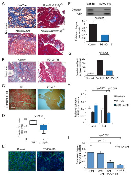
Figure 7. PI3Kγ promotes macrophage PDGF-BB expression to control PDAC fibrosis.
A. Masson’s Trichrome staining of tissue sections of pancreata from WT and p110γ−/− KC and KPC animals. Scale bar, 100μm. B. Masson’s Trichrome staining of sections of pancreata from control and TG100-115 treated KPC animals. C–D. Images (C) and quantification (D) of picrosirius red staining of sections of pancreata from KPC tumors grown in WT and p110γ−/− animals. Scale bar 100μm. E. Collagen I immunostaining of LMP tumors from animals treated with the PI3Kγ inhibitor TG100-115 or chemically similar inert control. F. Western blot and quantification of collagen I protein expression in LMP tumors from animals treated with the PI3Kγ inhibitor TG100-115 or chemically similar inert control. G. Relative collagen I mRNA expression in normal pancreata and LMP tumors from animals treated with the PI3Kγ inhibitor TG100-115 or chemically similar inert control. H. Relative collagen I mRNA expression in primary murine fibroblasts incubated in the presence or absence of stimulus free conditioned medium from IL-4 stimulated WT and p110γ−/− macrophages. I. Relative collagen I mRNA expression in fibroblasts incubated in the presence or absence of stimulus free conditioned medium from IL-4 stimulated WT macrophages in the absence or presence of anti-TGFβ, anti-PDGF-BB or Imatinib. Significance testing was performed by parametric Student’s t test.

