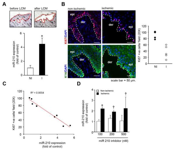
Figure 1.
(A) miR-210 expression from laser microdissected epidermis of human wound-edge tissue. (n=7), * p<0.001; ANOVA. (B) Serial human wound cross-sections stained with anti-Ki67 and keratin-14 antibody, counter stained with DAPI. (n=4-5). The plot represents quantification of the Ki67 positive cells/ field (20X). (C) Regression plot of miR-210 expression from the human wound edge biopsies against number of Ki67 positive cells/ field (20X). (n=8) (D) miR-210 expression from murine non-ischemic and ischemic wound-edge tissue 24h after intradermal delivery of naked LNA-anti-miR-210. (n=4). * p<0.01; † p<0.05 compared to nonischemic wound, ANOVA.

