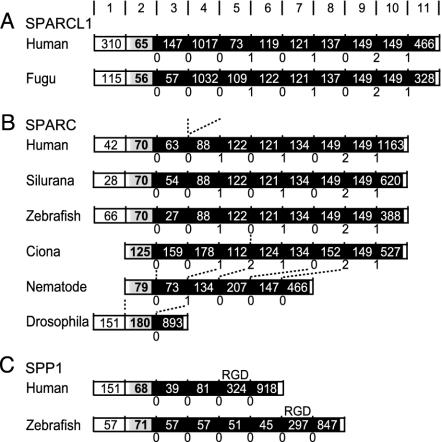
Fig. 2.
Structures of SPARCL1 (A), SPARC (B), and SPP1 (C). Boxes represent the untranslated region (white), signal peptide (localizes the protein in ECM; gray), and the mature protein (black). The length (nucleotide) of each exon is shown in the boxes. Intron phases are described below. Dashed lines show equivalent introns shifted by intron gain, loss, or sliding. (A) Exons 2–5 code domain I, which is separated by phase 0 introns. Exons 6 and 7 code domain II, and exons 8–11 code domain III. (B) Intron 4 in Ciona and intron 5 in nematode slide 1 base upward or downward, respectively. (C) The penultimate exon codes an Arg-Gly-Asp motif.

