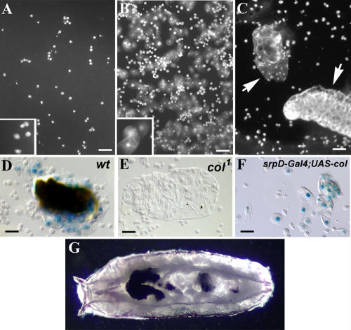
Figure 2. col Requirement for Lamellocyte Differentiation.
(A–C) 4′,6-diamidino-2-phenylindole (DAPI) staining of hemocytes from wt (A and B) and from col1 (C) third instar larvae. (A) Uninfected larva; (B) and (C) infected larvae. Plasmatocytes (inset in [A]) are always present, whereas lamellocytes (inset in [B]) are detected in the hemolymph of wt (B) but not col1 (C) larvae 48 h after infestation by L. boulardi. In col1 mutants, the wasp eggs are not encapsulated (white arrows) and develop into larvae (bottom right organism in [C]).
(D–F) Lamellocytes expressing the P-lacZ marker l(3)06949 (Braun et al. 1997) surround the wasp eggs in wt larvae (D), are completely absent in infected col1 mutant larvae (E), and differentiate in the absence of wasp infection following enforced Col expression in hematopoietic cells (srpD-Gal4/UAS-col larvae) (F). (G) srpD-Gal4/UAS-col pupa showing the presence of melanotic tumors.
Bars: 50 μm.

