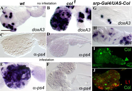
Figure 5. Col-Expressing Cells Play an Instructive Role in Lamellocyte Production.
Expression of the crystal cell marker doxA3 (Waltzer et al. 2003) (A, B, and G); of the lamellocyte markers α-ps4 (M. Meister, unpublished data) (C–F and H) and L1 (Asha et al. 2003) (J); and of Col (I and J); in wt (A, C, and E), col loss-of-function mutant (B, D, and F), and srp-Gal4/UAS-col (G–J) larvae. In (E) and (F), larvae were taken 48 h after infestation. An increased number of doxA3-positive cells (B) parallels the absence of lamellocyte differentiation (F) in col1 mutant lymph glands. Conversely, lamellocyte differentiation and a reduced number of doxA3-positive cells are observed upon enforced Col expression (G and H). Double staining for Col and L1 shows that Col-expressing cells and differentiating lamellocytes do not overlap in the lymph gland. (I) shows ectopic Col expression compared to expression in the PSC (arrowhead; not visible in [J]). Antibody and in situ probes are indicated on each panel. In all panels, larvae are oriented with the head to the left: a single primary lobe is shown, with sometimes a few secondary lobes. Bar: 50 μm.

