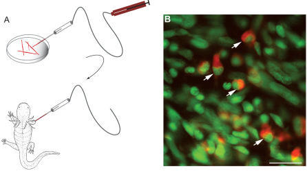
Figure 2. Cellularisation of Striated Myofibers after Implantation into a Larval Limb Blastema.
(A) Schematic diagram of procedure. After dissociation of larval limb musculature, the cells were loaded with a cell tracker dye and single myofibers taken up into a suction micropipette, prior to injection into a larval limb blastema as detailed in the Materials and Methods.
(B) Section of a limb at 48 h after implantation of CellTracker Orange-labelled myofibers. The section has been counterstained with the nuclear stain Sytox green. Note the dye-labelled mononucleate cells (arrowed). Scale bar, 20 μm.

